


NYCU Neuroscientists Illuminate the Brain’s Hidden “Star Map” of Neural Activity
(中央社訊息服務20251014 11:35:48)Researchers at the Institute of Neuroscience, National Yang Ming Chiao Tung University (NYCU), have developed a groundbreaking live imaging technology that can capture the electrical activity of neurons with unprecedented precision—an achievement recently published in Nature Methods under the title “Imaging neuronal voltage beyond the scattering limit.”
Much like pinpointing individual stars in a vast galaxy, this innovation overcomes one of neuroscience’s most significant observational barriers, marking a key advance in understanding how the brain truly works.

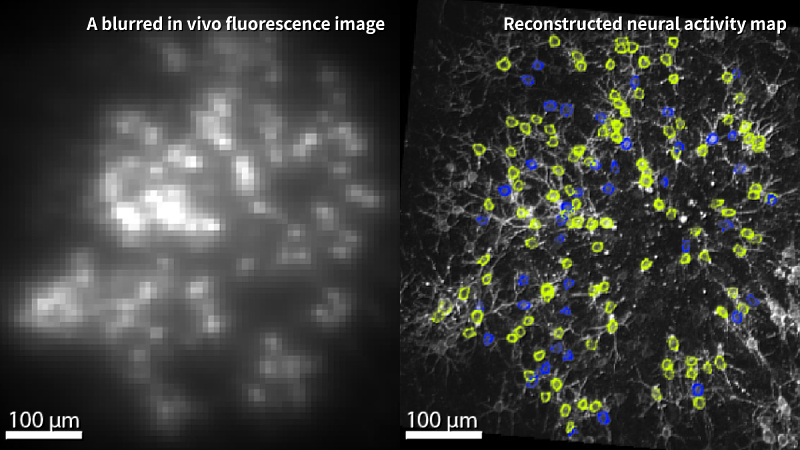
Our sensations, thoughts, and memories arise from lightning-fast electrical signals transmitted between neurons. Yet these signals often occur deep within the brain and vanish in milliseconds, making them nearly impossible to observe directly. Traditional optical imaging techniques struggle with light scattering, resulting in blurry halos rather than clear visualizations of neuronal activity.
Associate Professors Tsai-Wen Chen and Bei-Jung Lin addressed this challenge by utilizing voltage-sensitive fluorescent molecules to monitor subtle fluctuations in neuronal membrane potential. They discovered that while neural signals may overlap spatially, only a few neurons fire at any given moment. By treating these sparse flashes of electrical activity as positional clues—much like astronomers mapping the flicker of distant stars—the team achieved a new level of imaging clarity.
Their technique, termed Activity Localization Imaging (ALI), enabled the researchers to observe hippocampal neurons in live mice and pinpoint the exact coordinates of each neuronal discharge. By compiling tens of thousands of such events, they constructed a high-resolution “map” of neural activity.
“It’s like finding each shining star in the vast galaxy of the brain,” said Prof. Chen.
Last year, the same team utilized an earlier version of this technology to demonstrate that certain inhibitory neurons tend to fire in conjunction with specific groups of cells—a phenomenon reminiscent of social “friend groups” within the brain’s neural network.
Prof. Lin noted that this breakthrough now allows scientists to distinguish even smaller and more densely packed excitatory neurons, shedding new light on the neural circuits responsible for spatial cognition and memory formation. Although the current method cannot yet detect so-called “silent neurons” that do not actively fire, the researchers believe this represents a pivotal step toward visualizing brain activity at single-cell resolution in living organisms.
Led entirely by a Taiwan-based interdisciplinary team and involving international collaboration, this study demonstrates the strength and long-term investment of NYCU’s neuroscience research, marking a milestone in the nation’s contribution to global brain science.